
Product Description
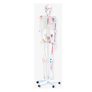
Model | BIX-A1002 — 180cm Human Skeleton Model with Colored Muscle & Ligament |
Summary | 180cm human skeleton with colored muscle and ligament: removable limbs, nerve branches, vertebral artery, lumbar disc; skull with movable jaw. PVC, washable. (157 chars) |
Height | 180 cm (full adult size) |
Highlighted Anatomy | Colored muscles & ligaments; nerve branches; vertebral artery; lumbar intervertebral disc |
Skull Detail | Movable jaw; removable cranium; epiphyseal line; three detachable lower teeth |
Limbs | Arms and legs removable |
Mounting | Metal bracket |
Material | PVC — washable, unbreakable; dust cover included |
Price | On request |
Scope of Review | Desk review based on published manufacturer specifications; confirm fine details with supplier |
Educational-use note: anatomy teaching aid — educational equipment, not a medical device or pharmaceutical product, and contains no medicinal ingredients.
1. What the A1002 Is (and What It Is Not)
The A1002 is the full-height flagship of the BIX skeleton line — a 180 cm adult skeleton that doubles as both an anatomy demonstrator and a room-scale display piece. Its specification sheet reads like a checklist for serious teaching:
Specification | Teaching Value |
Colored muscles & ligaments | Soft-tissue origins and relations visible on the bone |
Painted nerve branches | Nerve courses beyond what bare bone shows |
Vertebral artery display | Cervical vascular route on the actual skeleton |
Lumbar intervertebral disc | The pathology site of the world's most disabling condition — visible on the frame |
Removable arms & legs | Limb segments detach for station-based teaching |
Advanced skull | Movable jaw, removable cranium, epiphyseal line, 3 detachable lower teeth |
Metal bracket | Stable, permanent display mounting |
It is not a compact desktop skeleton (85 cm class) and not a lightweight portable trainer — it is the anatomy-hall instrument: the model students gather around, point at, and remember.
2. Why a Full-Height, Soft-Tissue-Enhanced Skeleton Earns Its Price
The clinical rationale for musculoskeletal anatomy education is difficult to overstate: musculoskeletal conditions are the leading contributor to disability worldwide, affecting an estimated 1.71 billion people, with low back pain the single leading cause of disability (WHO, 2022; GBD Low Back Pain Collaborators, 2023).
The teaching rationale is equally concrete. A systematic review and meta-analysis of randomized trials found that three-dimensional physical models were associated with significantly higher anatomy examination scores in medical students compared with 2-D methods (P<0.0001; Fleming et al., 2020) — consistent with the broad evidence base for model-enhanced instruction (Cook et al., 2011).
Where the A1002 differs from generic skeletons is in layering: it superimposes muscles, ligaments, nerves, and the lumbar disc onto the bone. That layering is exactly what novices struggle to reconstruct mentally — a painted origin on a full-height model is a fact they can see, not an abstraction they must memorize.
3. The Review Breakdown
3.1 Scale and Display Presence — ★★★★★
At full 180 cm adult height on a metal bracket, the A1002 carries a lecture or lab hall. This is the model that anchors an anatomy classroom, visible from any seat.
3.2 Soft-Tissue Color Coding — ★★★★★ (by spec)
Colored muscles and ligaments turn a skeleton into a musculoskeletal teaching surface. For programs teaching muscle origins, insertions, and joint relations, this is the single highest-value upgrade over a plain skeleton.
3.3 Spine, Nerve & Disc Detail — ★★★★★ (by spec)
Vertebral artery, nerve branches, and a lumbar intervertebral disc are displayed — structures normally invisible on bone. This gives the A1002 genuine neuro-vascular teaching capability that display skeletons usually lack.
3.4 Skull Detail — ★★★★★ (by spec)
A movable jaw, removable cranium, epiphyseal lines, and three detachable lower teeth make the skull a mini-teaching station in itself — including the epiphyseal-line anatomy that explains skull growth in the young.
3.5 Removability and Practicality — ★★★★☆
Arms and legs detach, enabling limb-level station teaching. At full height the model is a room piece, not a carry-around trainer — plan a fixed location.
3.6 Documentation Depth — ★★★☆☆
Core specifications are published (material, mounting, features), but fine details such as the exact number of painted muscles, articulation range, and replacement-part policy are not. Confirm with the supplier before ordering.
4. Model Positioning: A1002 in the Skeleton Line
Model | Height | Character | Best For |
A1002 | 180 cm | Colored muscle & ligament; nerves; vertebral artery; lumbar disc; advanced skull; removable limbs | Anatomy halls, flagship display & full-class teaching |
A1004 | 85 cm | Nerves + spine emphasis, green chest cartilage | Spine/neuroanatomy teaching stations |
A1006 | 42 cm | Compact skeleton | Individual desks, quick reference |
The line logic: A1002 shows everything at full scale; A1004 teaches the spine and nerves specifically; A1006 puts a skeleton on every desk.
5. Verified Strengths & Honest Limitations
Strengths
● Full adult scale with soft-tissue color coding — rare in one product.
● Neuro-vascular and disc display (vertebral artery, nerve branches, lumbar disc) adds teaching depth beyond bone.
● Advanced skull details (movable jaw, removable cranium, epiphyseal line, detachable teeth) for craniofacial and growth teaching.
● Removable limbs support station-based instruction.
● PVC construction: washable and unbreakable; dust cover included — practical for high-traffic halls.
Limitations / Points to Confirm
● Full-height = fixed-location display; not a portable trainer.
● Painted muscle/ligament display is schematic (educational color coding) — not a substitute for cadaveric or digital detail at muscle level.
● Exact painted-muscle count, articulation range, and parts policy are unpublished — request the data sheet.
● Price on request; MOQ, packaging, and freight to be confirmed.
6. FAQ
Q1: What is the difference between A1002, A1004, and A1006? A: A1002 (180 cm) is the full-height flagship with colored muscles/ligaments, neuro-vascular detail, and an advanced skull; A1004 (85 cm) is the mid-size spine-and-nerves teaching model; A1006 (42 cm) is the compact reference skeleton. Choose by hall space and teaching depth.
Q2: Are the muscle and ligament colors anatomically accurate? A: The color coding is an educational display of muscle and ligament structures on the bone — schematic by design, not a substitute for cadaveric dissection. It is excellent for origins/relations teaching; confirm the detail level you need with the supplier.
Q3: What can the skull teach? A: The movable jaw demonstrates mastication mechanics; the removable cranium and epiphyseal lines show skull structure and growth; the detachable lower teeth support dental anatomy teaching.
Q4: Why are the arms and legs removable? A: Detachable limbs allow the model to be split into teaching stations (upper limb, lower limb) and simplify transport of a full-size skeleton.
Q5: How durable is it, and how do I clean it? A: PVC construction is washable and unbreakable, and a dust cover is included. Wipe with a damp cloth; store covered when not in use.
Q6: What should I confirm before ordering? A: Painted-detail coverage, mounting options, replacement parts, packaging, and freight. Email cpr480@adaanatomy.com — we will send the specification sheet and a quote.
References
Musculoskeletal Health — WHO Fact Sheet (2022)
Global, Regional, and National Burden of Low Back Pain, 1990–2020 — GBD Low Back Pain Collaborators (2023), Lancet Rheumatol
Effectiveness of Three-Dimensionally Printed Models in Anatomy Education for Medical Students and Resident Physicians: Systematic Review and Meta-Analysis — Fleming et al. (2020), J Am Coll Radiol 17(10):1220–1229
Technology-Enhanced Simulation for Health Professions Education: A Systematic Review and Meta-analysis — Cook et al. (2011), JAMA 306(9):978–988