
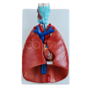
Reading the Bronchial Tree: How a Transparent Heart-Lung Model Changes Respiratory Teaching
Students can recite that the right lung has ten segments and the left has eight, and still fail to say where a bronchoscope is pointing. The gap is spatial, and it closes when the bronchial tree is visible through the lung surface. The BIX-A1072 is built for that: a throat, heart and lung model at 2× magnification with transparent lung shells, showing the trachea and bronchial tree in plastic, and segment structure of eight on the left and ten on the right. It is listed at US $69.24.

Quick facts
| Item | Detail |
|---|---|
| Model | BIX-A1072 |
| Scope | Throat, heart and lungs as a unit |
| Magnification | 2× |
| Lung shells | Transparent plastic, showing segment boundaries |
| Airway | Trachea and bronchial tree modelled in plastic |
| Segmentation | Right lung 10 segments, left lung 8 segments (per listing) |
| Listed price | US $69.24 |
| Typical use | Anatomy courses, thoracic surgery teaching, anaesthesia and respiratory therapy |
The teaching problem the model solves
The bronchial tree is a three-dimensional branching structure taught with two-dimensional diagrams. That mismatch produces a predictable error pattern: students learn the names of the lobar bronchi but cannot predict where a segmental bronchus will appear on bronchoscopy, and they cannot relate a chest X-ray opacity to a segment.
A transparent model changes the question from "name this branch" to "find this branch", which is the question asked in the skills lab. Radiology-based anatomy teaching studies show measurable learning gains when cross-sectional imaging is integrated with anatomical teaching [2]; a transparent airway model provides the physical counterpart to that imaging view.
For anaesthesia and critical care, the stakes are practical. Lung isolation — placing a double-lumen tube or bronchial blocker correctly — depends on recognising bronchial anatomy through a fibreoptic view. A study of novices learning basic lung isolation skills on an anatomy simulator found that trainees could be brought to a basic proficiency level, and that skills decayed over time without practice [1]. Visibility of the tree during training is what makes that proficiency achievable in a short course.
Three drills that use the bronchial tree
Drill 1 — Segment mapping (10 min). Students trace the trachea to each lobar bronchus and then to named segments, marking their route on a worksheet. The transparent shell lets the instructor check the route without disassembly.
Drill 2 — Imaging correlation (15 min). Show a chest CT slice at the level of the carina and ask students to identify the corresponding structures on the model. Repeat at the level of the middle lobe bronchus.
Drill 3 — Tube placement reasoning (15 min). Without a bronchoscope, ask students to explain where a left-sided double-lumen tube should sit and why the left main bronchus is usually chosen. Then demonstrate the anatomy that makes the answer true.
Heart and throat in the same unit
Including the heart and upper airway makes the model useful beyond respiration. The relationship between the trachea, oesophagus and aortic arch explains why an enlarging aortic aneurysm can cause tracheal deviation, and why an oesophageal foreign body can compress the airway. Those correlations are difficult to teach from separate organ models and straightforward on a combined unit.
At 2× magnification, the model is large enough for group demonstration — approximately the size of a desktop teaching unit — while remaining portable between classrooms.
Limits worth knowing
It is a static anatomical model, not a functional simulator: there is no airflow, no compliance, no bronchoscope channel. For bronchoscopy technique, you need an airway trainer with a working lumen.
The transparent shell also shows wear: repeated handling dulls the surface and reduces visibility. Store it covered and clean with a soft cloth rather than solvent.
Procurement notes
- Confirm segmentation — the listing specifies 10 right and 8 left segments; verify in writing if your curriculum uses a different convention.
- Check the magnification and footprint — 2× magnification implies a desktop unit; confirm dimensions fit your demonstration bench.
- Ask about the stand — a combined heart-lung unit needs a stable base; confirm inclusion.
- Request the segment key — a printed legend speeds up Drill 1 and reduces instructor correction time.
- Plan storage — dust is the main enemy of transparency; ask whether a cover is available.
FAQ
Q1. How many lung segments does the model show?
Eight on the left and ten on the right, as specified in the product listing.
Q2. What is the magnification and price?
2× magnification; listed at US $69.24 per unit.
Q3. Can it be used for bronchoscopy training?
No — it is an anatomical model. Bronchoscopy requires an airway trainer with a working channel; this model supports the anatomy teaching that precedes it.
Q4. Is it suitable for anaesthesia trainees?
Yes for airway and segment anatomy. Pair it with a lung-isolation trainer for the procedural component [1].
Q5. Does it include the heart?
Yes — the unit covers throat, heart and lungs together, which supports cardiopulmonary correlation teaching.
Q6. How do we request a quotation?
Email cpr480@adaanatomy.com with the quantity, your city and whether you need the segment key printed.
References
- Woo JA, et al. Teaching basic lung isolation skills on human anatomy simulator: attainment and retention of lung isolation skills. BMC Anesthesiology. 2016. PMID 26790624.
- Kournoutas I, et al. The educational impact of radiology in anatomy teaching: a field study using cross-sectional imaging. Academic Radiology. 2024. PMID 37925345.
Contact
For the specification sheet, segment key and a landed-cost quotation: cpr480@adaanatomy.com. Production follows ISO 45001 management practice; CE documentation available for EU procurement.