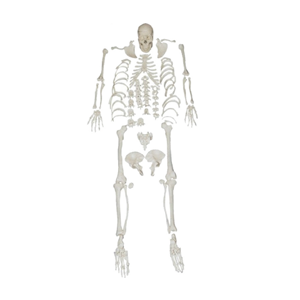
The whole-body bone scattering model is a professional model often used in medical education, anatomical research and related teaching demonstrations. It is composed of all the major bones of the human body, including the skull, spine, ribs, sternum, pelvis, upper and lower limb bones, etc. Each part is precisely fabricated in accordance with anatomical proportions and structures, and can be disassembled and reassembled independently. The most distinctive feature of this type of model is that the bones are scattered and independent. Users can clearly observe the shape of each bone, the connection mode of the joints, and the spatial relationship between them. By manual disassembly and assembly, learners can not only have an intuitive understanding of the overall structure of the human skeleton, but also deeply master the anatomical details of individual bones.
In medical teaching, the whole-body bone scattering model helps medical students, nursing professionals and rehabilitation trainees establish a systematic concept of bones, strengthen memory and improve practical operation skills. At the same time, it can also provide references for forensic medicine, kinesiology and art modeling. For instance, when forensic experts conduct bone appraisals, they can use this model for comparison. In the creation of human body sculptures, artists often use bone-scattering models as anatomical references. Its materials are mostly made of high-strength plastics or resins, balancing durability and realism. Some high-end versions even feature bone texture and weight simulation. Overall, the whole-body bone scattering model is not only an ideal tool for anatomy teaching but also an important auxiliary device in multiple disciplinary fields.