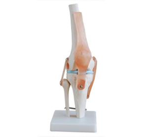
The natural large knee joint model recreates the femur, tibia, fibula and patella at a 1:1 scale, marking the articular surface, cartilage and meniscus, and color matching shows the anterior and posterior cruciate ligaments and the medial and lateral collateral ligaments. The joints can perform flexion, extension and slight rotation, demonstrating the "lock-unlock" mechanism, which is suitable for teaching anatomy, sports medicine and rehabilitation.
The models are mostly made of durable PVC and elastic silicone, partially transparent, and the ligaments can be disassembled and replaced, which is convenient for explaining the injury and surgical repair path. The magnetic or bolted base is stable, portable and easy to store. The accompanying pathological parts can simulate meniscus tears, bone spurs and cartilage wear, assisting in preoperative communication and functional assessment.
It is recommended to use a ruler for Angle measurement and a muscle strength tape for teaching when in use to enhance interaction. For maintenance, wipe with a slightly damp cloth, avoid strong light, high temperature and alcohol corrosion. Regularly check the movable shaft and fasteners to ensure smooth damping and accurate teaching.
The model can demonstrate the bending, extension and inward/outward rotation of the knee joint. Naturally large and with a base.