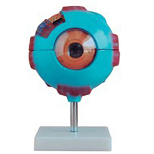
The eyeball magnification model is a commonly used anatomical model for teaching and research. By proportionally magnifying the structure of the human eye, it enables learners to clearly observe the complex internal structure of the eyeball.
Models typically display key parts such as the cornea, iris, pupil, lens, vitreous body, retina and optic nerve, and can be disassembled to help understand the spatial relationships and functions of each part. This model holds significant value in the teaching of medicine, nursing, biology and visual science. It can visually demonstrate the principles of light refraction and imaging, and assist in explaining the formation mechanisms of common ophthalmic diseases such as cataracts and glaucoma.
Through the demonstration of the eyeball magnification model, students can master the structure and physiological functions of the eye more accurately, laying a solid foundation for subsequent clinical operations and disease diagnosis.