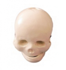
The main function of infant skull models lies in assisting medical teaching and clinical skills training. Because the infant's skull has not yet fully ossified, the cranial sutures and fontanelles are soft and have not closed. Therefore, understanding its structure and developmental characteristics is particularly important for medical staff in pediatrics, obstetrics, neurology and other fields. Through the infant skull model, learners can intuitively grasp the anatomical structure of the infant's skull, including the positions and shapes of the frontal bone, parietal bone, occipital bone and fontanelle, thereby understanding the changing patterns of the infant's head during its growth and development.
In addition, the model can also be used to demonstrate the deformation of the fetal head and the mechanism of its passage through the birth canal during childbirth, helping midwives and medical students master safe delivery techniques. It also plays a significant role in neonatal care teaching and can be used to train how to correctly touch the fontanelle, identify abnormal cranial shapes, and assess changes in intracranial pressure. Through the operation and observation of the model, students can not only enhance their theoretical knowledge but also improve their clinical judgment and practical abilities. Overall, the infant skull model is an indispensable teaching tool in medical education, providing intuitive and effective support for improving the clinical level of pediatrics and obstetrics.