The standard operation of the natural large skull model is mainly used in medical teaching, oral anatomy, surgical training and forensic research, which can help learners intuitively grasp the structure and spatial relationship of the skull.
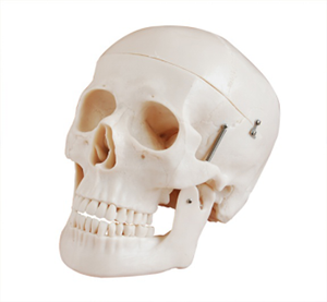
Before operation, it is necessary to first check whether the model is complete and confirm that all parts of the skull, such as the craniocalyx, zygomatic bone, mandible, etc., are firmly connected. When in use, first, according to the teaching requirements, open the skull cover of the skull model to display the internal structure of the cranial cavity, including the frontal bone, parietal bone, occipital bone and the shape of the brain cavity.
Secondly, the mandible can be disassembled and repositioned to help trainees understand the movement mode of the temporomandibular joint and the arrangement of the dental arch. For models with markings, one can also learn the various anatomical details of the skull, such as the positions of the orbital cavity, nasal cavity, and skull base foramen, by referring to the numbers, which is convenient for remembering the entry and exit channels of nerves and blood vessels.
During operation, handle with care to avoid damage to joints or weak areas. After use, all parts should be restored and placed in a stand or storage box to keep it clean and dry. Through standardized operations, learners can master the knowledge of cranial anatomy more systematically, enhance their spatial imagination and clinical application ability.